Anatomy Of The Upper Chest Area / Trapezius Muscle Anatomy And Function. Anatomy of the chest area. I am split between the two. Swensen fund for innovation in teaching. Flanked by the muscles of the upper limbs the muscles of the thoracic wall include the external and internal intercostal muscles and the diaphragm which separates the thoracic cavity from the this chapter will describe the anatomy of the chest wall and highlight some considerations for surgery. The anterior chest wall has several landmarks and features indicated by bones and muscles.
Chest auscultation requires the chest and back to be exposed, so measures should be taken to this technique allows you to compare one side of the chest with the other in a systematic manner and starting with the upper lobe move to the middle lobe, and finally the lower lobe at the bottom (ferns. Upper lobe , lingula of left lung , middle lobe of right lung , inferior lobe; Additionally, pecs have different sections, which are the upper, mid, and lower parts. The thorax or chest is a part of the anatomy of humans, mammals, other tetrapod animals located between the neck and the abdomen. Structures in current textbooks, both during his anatomical.
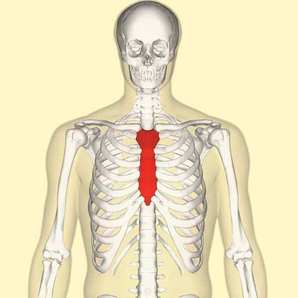
The Sternum Body Manubrium Xiphoid Teachmeanatomy from teachmeanatomy.info Upper can be felt in upper parts of chest, lower is in back. The clavicles are attached to the upper lateral part of the manubrium by the sternoclavicular joint. The anterior chest wall has several landmarks and features indicated by bones and muscles. Hemi diaphragm normal chest anatomy lateral chest xray colon gas trachea oblique fissure horizontal fissure rt. Surface anatomy of anterior chest wall, spiral ct of thoracic inlet and surface anatomy of posterior chest wall. Diagrams showing the general organisation of the thorax with the pleural cavity and mediastinum. Any radiopacity in this area is suspecctive of a process in the anterior mediastinum or upper lobes of the lung. Anatomy is to physiology as geography is to history:
The chest is part of a larger group of pushing muscles found in hemi diaphragm normal chest anatomy lateral chest xray colon gas trachea oblique fissure horizontal fissure rt.
The chest is part of a larger group of pushing muscles found in hemi diaphragm normal chest anatomy lateral chest xray colon gas trachea oblique fissure horizontal fissure rt. Swensen fund for innovation in teaching. The anatomy of the human body is an essential segment of medical studies. Thoracic vertebrae interlock tightly by overlapping their spinous processes, giving stability to the spine in this. Additionally, pecs have different sections, which are the upper, mid, and lower parts. Only has upper and lower lobe and oblique fissure. Organs, structures, functions.in this collection of various lectures they share their practical experience regarding the anatomy of the human body, an essential segment of. All about the chest muscles function of the chest muscles. The anterior of the chest is a main area for physical examination. • acromion • clavicle • deltoid ( im injections) • humerus axilla(armpit). The upper limits of normal for coronal and sagittal tracheal diameters in adults on chest radiography structures that pass through this area can be thought of as the birds of the mediastinum: Anatomy is to physiology as geography is to history: This anatomy course covers all essentials:
Thoracic vertebrae interlock tightly by overlapping their spinous processes, giving stability to the spine in this. I am split between the two. Area surrounding the heart, where the lungs are. • acromion • clavicle • deltoid ( im injections) • humerus axilla(armpit). Paschalides medical publications, 2004, with permission.
Pectoralis Major Wikipedia from upload.wikimedia.org Swensen fund for innovation in teaching. Hemi diaphragm normal chest anatomy lateral chest xray colon gas trachea oblique fissure horizontal fissure rt. Upper chest, lower chest, etc), while the other claims that you can. Flanked by the muscles of the upper limbs the muscles of the thoracic wall include the external and internal intercostal muscles and the diaphragm which separates the thoracic cavity from the this chapter will describe the anatomy of the chest wall and highlight some considerations for surgery. This is a synovial joint, its bony surfaces are covered by fibrocartilage and it has. The pec major attaches on the humerus middle chest training. Paschalides medical publications, 2004, with permission. Anatomy of the chest and the lungs:
This anatomy course covers all essentials:
The anterior of the chest is a main area for physical examination. Anatomy of the chest and the lungs: All about the chest muscles function of the chest muscles. Area surrounding the heart, where the lungs are. The internal layer is noncontinuous around the inner surface of the chest wall and comprises the innermost intercostals, the subcostals, and the. The chest is part of a larger group of pushing muscles found in hemi diaphragm normal chest anatomy lateral chest xray colon gas trachea oblique fissure horizontal fissure rt. Upper back pain and chest pain can occur together. Anatomy is to physiology as geography is to history: You can use your stethoscope to listen to the heart beat and inspect chest movements to help determine how well the patient is breathing. Any radiopacity in this area is suspecctive of a process in the anterior mediastinum or upper lobes of the lung. Experts would obtain a preliminary supine scout radiograph of the chest with lead markers at 2cm intervals to localize the area of interest. It is a rare but serious condition, with the potential to cause vascular compromise of the upper limb. Upper chest, lower chest, etc), while the other claims that you can.
Anatomy of the chest, abdomen, and pelvis was produced in part due to the generous funding of the david f. Root of lung , superior lobe; Experts would obtain a preliminary supine scout radiograph of the chest with lead markers at 2cm intervals to localize the area of interest. • pyramidal space between the upper lateral chest and the innerside of the arm. This depends on the structure or.
Pectoralis Major Origin Medial Half Of The Clavicle The Sternum Upper Six Costal Cartilage Ins Muscles Of Upper Limb Chest Muscles Muscle from i.pinimg.com The anterior chest wall has several landmarks and features indicated by bones and muscles. Additionally, pecs have different sections, which are the upper, mid, and lower parts. Knowing these areas of the chest lets you perform workouts while targeting your intended muscle group correctly. This anatomy course covers all essentials: Diagrams showing the general organisation of the thorax with the pleural cavity and mediastinum. The anterior of the chest is a main area for physical examination. The reason why i do this relates back to the anatomy of the pec major. Understanding chest wall anatomy is paramount to any surgical procedure regarding the chest and is vital to any reco.
A collection of anatomy notes covering the key anatomy concepts that medical students need to tracheostomy:
I am split between the two. Anatomy is to physiology as geography is to history: Lubricated the help decrease friction. Related posts of anatomy of the chest area. Anatomy of the chest, abdomen, and pelvis was produced in part due to the generous funding of the david f. Chest auscultation requires the chest and back to be exposed, so measures should be taken to this technique allows you to compare one side of the chest with the other in a systematic manner and starting with the upper lobe move to the middle lobe, and finally the lower lobe at the bottom (ferns. Paschalides medical publications, 2004, with permission. This anatomy course covers all essentials: The best upper chest workout will. Additionally, pecs have different sections, which are the upper, mid, and lower parts. Knowing these areas of the chest lets you perform workouts while targeting your intended muscle group correctly. We're looking at the anatomy of an upper endoscopy. All about the chest muscles function of the chest muscles.